This review article discusses the various analytical techniques for the characterisation of Polymeric Nanoparticles (PNPs). Because of their distinctive qualities, they have gained a lot of attention in recent years. As a result, knowing their properties is important. In fact, a range of analytical techniques are used to investigate these features. It includes microscopic, spectroscopic, light scattering, and diffraction techniques. This article will provide you with a review of these characterisation methods and how they are used to study the properties of PNPs.
Rosemary Salin, Cademix Institute of Technology
Introduction
Nanotechnology is the development of molecular-scale functional systems. It emerged as a field in the 1980s. Later on, this sector found great demand in fields such as medicine, electronics, energy storage, cosmetics, and so on. Nanoparticles (NPs) are a broad category of materials with at least one dimension smaller than 100 nanometers. When scientists discovered that size may affect a substance’s physiochemical properties, they realised the importance of these materials. 1 Depending on their physical and chemical properties, there are different types of NPs. They are: carbon-based NPs, metal NPs, ceramic NPs, semiconductor NPs, lipid-based NPs and polymeric NPs. 1, 2
In recent years, Polymeric nanoparticles (PNPs) are popular due to their wide variety of uses. For example, it has applications in photonics, electronics, sensing, medicine, pollution control, and environmental applications. In fact, PNPs are most commonly used in drug delivery and medicine. 3 Different characterisation methods help in the analysis of these properties. Firstly, let us discuss about PNPs and some of their methods of preparation.
What are Polymeric Nanonoparticles (PNPs)?
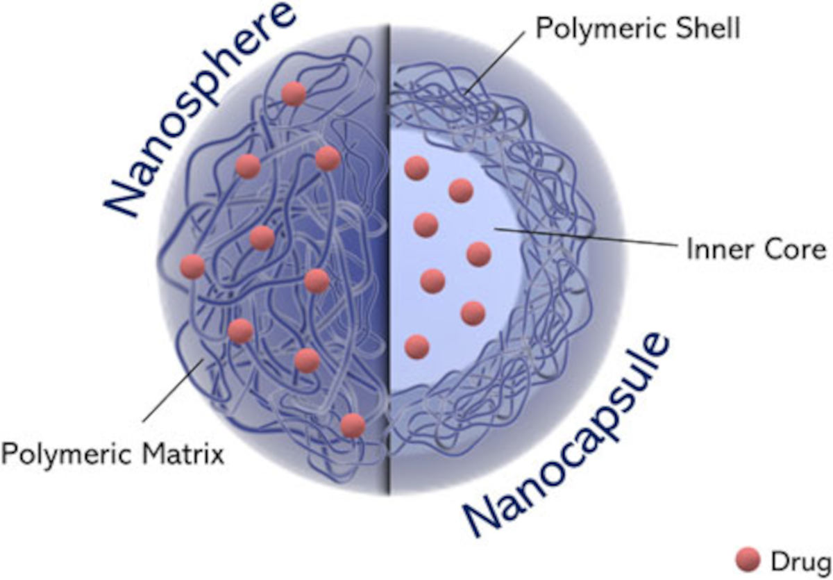
Polymeric nanoparticles are organic NPs. 1 Typically, they are in 1 to 1000 nm size range. 1, 5, 6, 7 They are prepared from biocompatible and biodegradable polymers. In addition, we can load active substances into them. The polymeric core can either entrap these active substances or adsorb them onto the surface. Generally, they are nanospheres or nanocapsules. Nanospheres are matrix-type NPs where the entire mass is solid and consists of spherical polymeric matrices. 1, 5 Moreover, in them, an even distribution of the active chemicals occurs throughout the polymeric core. Besides, drugs can adsorb on the surface of nanospheres. A nanocapsule, on the other hand, is a nanoscale shell. It has a space to insert the active substances. Figure 1 4 depicts the distinction between these two polymeric NPs.
Because of their nanosize, polymer nanoparticles have properties that the macroscale polymer does not have. This conversion from bulk polymer to nano polymer brings new properties. However, the composition of the material will not change. 3 These qualities have led to their application as drug delivery agents or nanocarriers. Despite the fact that the focus of this paper is on techniques for characterising polymeric nanoparticles, let us look at their synthesis methods.
Methods for Preparation of Polymeric Nanoparticles
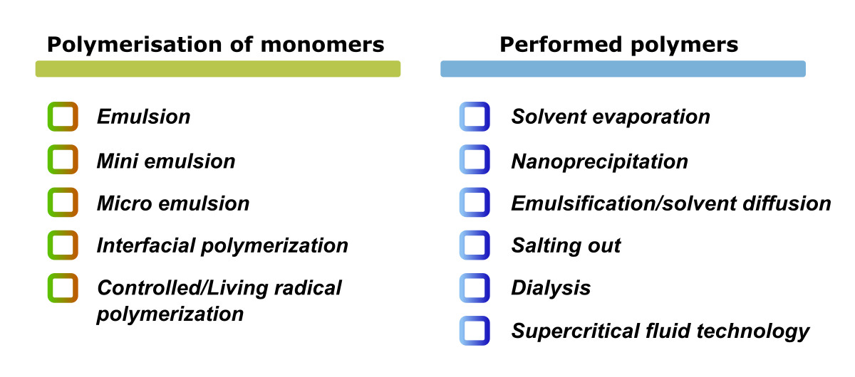
There are a variety of methods for the synthesis of polymeric nanoparticles depending on their use and the physicochemical properties of the drug. The selection of the most appropriate method is critical in order to get PNPs with the desired properties for a certain application. 2, 5, 7–10 The preparation methods are classified into two groups. They are; those based on monomer polymerisation and those utilising preformed polymers 2, 5–9 (Figure 2).
Characterisation Methods of Polymeric Nanoparticles
Polymeric NPs can differ in physical parameters such as composition and concentration, as well as size, shape, surface properties, crystallinity, and dispersion state. There are several analytical methods to analyse these properties. These methods include microscopic, spectroscopic, diffraction, and light scattering techniques. 2, 5, 8,10
Microscopic Techniques
Transmission Electron Microscopy (TEM)
Conventional light microscopy has difficulty resolving nanoscale structures. However, electron microscopic techniques can provide fine features of these nanostructures. Transmission electron microscopy (TEM) and Scanning electron microscopy (SEM) are the most commonly used electron microscopic techniques. Both of them will give same kind of information.
In TEM, passing an electron beam through a specimen results in the formation of the image. In other words, the interaction of the electrons with the sample produces an image. After that, the picture is magnified and focussed onto an imaging device. Thus the data is collected. Generally, TEM can distinguish between nanocapsules and nanospheres. In addition, we can get the thickness of the nanocapsule wall using TEM. Thus, it is an effective method for determining the shape and size of nanoparticles. 2, 5, 8,10
Scanning Electron Microscopy (SEM)
SEM is a type of electron microscope that scans the surface of a sample with a concentrated stream of electrons to obtain images of the sample. When electrons contact with atoms in a sample, they produce a variety of signals that carry information about the sample’s surface topography and composition. 2 SEM is also a method which helps in the determination of the size, size distribution and morphology of polymeric nanoparticles. Besides, it can give the degree of aggregation of PNPs. 5, 8
For example, Anwer et al 11 developed PLGA polymeric nanoparticles by single emulsion and evaporation technique. They studied at the potential of Eudragit S100-coated PLGA-based nanoparticles to entrap and release ELUX in a controlled manner for colon targeted drug delivery. They also recorded the SEM images for ELUX loaded plain (without Eudragit coating) PLGA NPs and coated NPs (Figure 3). 11 According to the SEM images, both plain and coated NPs were spherical in form and had a smooth surface with some aggregation. 11
![SEM images of (A) plain and (B) coated PLGA Polymeric nanoparticles from article [11]](https://www.cademix.org/wp-content/uploads/sem-images-rosemary-cademix.jpeg)
Atomic Force Microscopy (AFM)
AFM is another technique that has been used to characterise the surface morphology of polymeric NPs (AFM). Scanning probe microscopy is another name for it (SPM). It is useful not only for studying surface morphology with nanoscale precision, but also for measuring sensitive forces. 5, 8
An AFM scans across a sample surface with the sharp tip of the cantilever. As the tip reaches the surface, the cantilever deflects towards the surface due to the attractive force between the surface and the tip. However, when the cantilever gets closer to the surface and the tip makes contact with it, repulsive force takes control and causes the cantilever to deflect away from the surface. 5 A laser beam detects cantilever deflections towards or away from the surface. As a result, any cantilever deflection will create tiny variations in the direction of the reflected beam. These changes may be tracked using a position-sensitive photo diode (PSPD). 5
Light Scattering Techniques for Polymeric nanoparticles
The detection of light scattered by the interaction of light with matter provides information about the physical properties of the sample. In light scattering studies, firstly, a monochromatic beam is aimed at the sample. After that, the scattered light is recorded at a certain angle by a detector. Thus the properties of the sample are studied. There are three basic light scattering techniques used for characterising polymeric nanoparticles.
Dynamic Light Scattering (DLS)
Dynamic light scattering (DLS) or photon correlation spectroscopy is another method for the particle size determination. 2, 5, 8 DLS calculates the size of nanoparticles in a solvent. It is based on the Brownian motion of the particles. 2, 5, 9 Firstly, a DLS experiment involves the irradiation of a monochromatic laser light to a colloidal solution. Then the light is then scattered into a photo detector. The scattered light intensity changes in time due to the Brownian motion of the particles. This fluctuation in light intensity with time is connected to the particle size using an autocorrelation function. As a result, particle size determination is possible using DLS. DLS measurements are appropriate for monodisperse and polydisperse materials, but not for high-dimensional samples. It also allows for the calculation of the polydispersity index, which indicates the heterogeneity of NP size in a suspension. 9
Electrophoretic Light Scattering (ELS)
Dynamic light scattering is the foundation of electrophoretic light scattering. DLS helps in particle size determination whereas, ELS helps to compute zeta potential by measuring electrophoretic mobility. It is the best method for zeta potential determination. In fact, zeta potential is a measure of stability of colloidal solutions. It also helps in the analysis of surface properties. 5 Therefore, values between – 30 mV and +30 mV suggest instability, aggregation, coagulation, or flocculation. As a result of electric repulsion, particle aggregation is less at high zeta potential values. 5
Spectroscopic Techniques
Spectroscopic techniques provides a lot of information about the chemical structure and properties of PNPs. So, let us see some of the commonly used spectroscopic techniques for the characterisation of polymeric nanoparticles.
Fourier transform infrared spectroscopy (FT-IR)
Fourier transformed infrared spectroscopy (FTIR) is a spectroscopic method that measures vibrational transitions between distinct molecular excitation states. 5, 10 This method is beneficial for studying the structural features of PNPs. Moreover, it is also useful to study the molecular interactions between medicines and the encapsulating polymer. In fact, we can identify the presence of known compounds in the fingerprint region. In addition, we can detect the presence of impurities. 2, 5 Anwer et al. 9 for instance, used FTIR spectroscopy to study the interaction between ELUX drug and the PLGA NPs.
UV-Visible Spectroscopy
Nanoparticles have unique optical properties. These properties are sensitive to size, shape, concentration, and agglomeration of NPs. 10 This makes UV-Visible spectroscopy a suitable method for detection, and characterisation of polymeric nanoparticles. It is a spectroscopic technique that measures analyte concentration based on the amount of light absorbed by the sample. It has also been used to analyse the conjugation and conjugation ratio of biomolecules to nanomedicines. For example, Qasim et al. studied the encapsulation of silver nanoparticles in poly-N-isopropylacrylamide-based polymeric nanoparticles using UV-Visible spectroscopy. 12
X-ray Photoelectron Spectroscopy (XPS)
X-ray photoelectron spectroscopy (XPS) is a method for evaluating the surface chemistry of a material. It can determine the elemental composition of a material as well as its chemical and electronic states. 5 In XPS, firstly, a solid surface is irradiated with an X-ray beam. As a result, excitation of electrons to discrete energy levels occurs. Then the kinetic energy of electrons released from the top 1-10 nm of the material is measured. It will help in determining the elemental composition.
Fluorescence Spectroscopy
Fluorescence spectroscopy is a spectroscopy technique that studies fluorescence from a sample. Fluorimetry is another name for it. It is utilising a beam of light, generally ultraviolet light, to excite the electrons in a particular compounds. Consequently, these compounds will emit light. 10 This analytical method has a variety of applications. For example, it is used to study biomaterial conformational changes and their conjugation with nanoparticles. Furthermore, other researchers used the fluorescence abilities of the drug to assess its encapsulation efficiency in PNPs and its release. 10
In addition to the above methods, Nuclear Magnetic Resonance (NMR), and Mass spectrometry are also helpful for studying the structure, composition, and molecular weight.
Diffraction Techniques
Diffraction techniques are useful for studying the crystal structure of the sample. Generally, XRD analysis is used for this purpose. So, let us see the X-ray diffraction technique used for crystal structure determination of polymeric nanoparticles.
X-ray Diffraction (XRD)
X-ray diffraction (XRD) is an useful method for studying the crystal structure of NPs. It is used for the main characterization of material properties such as crystal structure, crystallite size, and strain. 2 In fact, depending on the structure of the crystal and the wavelength of the light interacting with the crystal, different diffraction patterns appear. The interference of reflected rays from different layers of the material in the periodic structure causes diffraction. 2
Conclusion
Polymeric nanoparticles have several uses. One of the most important use is in nanomedicine. As a result, knowing the properties of PNPs is highly important. The properties of PNPs are studied using a variety of analytical methods. It includes methods like as microscopy, spectroscopy, diffraction, and light scattering techniques. This review article discusses some important analytical techniques in PNP characterisation. Generally, microscopic techniques gives details like size, size distribution, and agglomeration of NPs. Similarly, DLS is also useful for particle size determination. However, spectroscopic techniques can help to find structural details and composition. Moreover, XRD technique helps to study the crystal structure of crystalline NPs.
Biblography
1. Khan, I., Saeed, K. & Khan, I. Nanoparticles : Properties, applications and toxicities. Arab. J. Chem. 12, 908–931 (2019).
2. Aslı Katmıs, Serap Fide, Seyma Karaismailoglu, S. D. Synthesis and characterization methods of polymeric nanoparticles. 2, 1–9 (2019).
3. Adhikari, C. Polymer nanoparticles-preparations, applications and future insights: a concise review. Polym. Technol. Mater. 60, 1996–2024 (2021).
4. Gagliardi Agnese, Giuliano Elena, Venkateswararao Eeda, Fresta Massimo, Bulotta Stefania, Awasthi Vibhudutta, C. D. Biodegradable Polymeric Nanoparticles for Drug Delivery to Solid Tumors. Front. Pharmacol. 12, (2021).
5. Carina I.C. Crucho, M. T. B. Polymeric nanoparticles : A study on the preparation variables and characterization methods. Mater. Sci. Eng. C 80, 771–784 (2017).
6. Nagavarma, B. V. N., Yadav, H. K. S., Ayaz, A., Vasudha, L. S. & Shivakumar, H. G. Different techniques for preparation of polymeric nanoparticles- A review. Asian J. Pharm. Clin. Res. 5, 16–23 (2012).
7. Jones, S. A., Mesgarpour, S., Chana, J. & Forbes, B. Preparation and Characterisation of Polymeric Nanoparticles Using Low Molecular Weight Poly ( vinyl alcohol ). J. Biomed. Nanotechnol. 4, 319–325 (2008).
8. Zielińska, A. et al. Polymeric Nanoparticles: Production, Characterization, Toxicology and Ecotoxicology. Molecules 25, 3731 (2020).
9. Ramalho, M. J. & Pereira, M. C. Preparation and Characterization of Polymeric Nanoparticles: An Interdisciplinary Experiment. Journel Chem. Educ. 93, 1446–1451 (2016).
10. Fornaguera, C. & Solans, C. Analytical Methods to Characterize and Purify Polymeric Nanoparticles. Int. J. Polym. Sci. 2018, (2018).
11. Anwer Md. K., Al-Shdefat Ramadan, Ezzeldin Essam, Alshahrani Saad M., Alshetaili Abdullah S., I. M. Preparation , Evaluation and Bioavailability Studies of Eudragit Coated PLGA Nanoparticles for Sustained Release of Eluxadoline for the Treatment of Irritable Bowel Syndrome. Front. Pharmacol. 8, (2017).
12. Qasim M, Udomluck N, Chang J, Park H, K. K. Antimicrobial activity of silver nanoparticles encapsulated in poly-N-isopropylacrylamide-based polymeric nanoparticles. Int. J. Nanomedicine 13, 235–249 (2018).
About the Author
Rosemary Salin is an Associate Chemist at Cademix Institute of Technology, Austria with a Master of Science in Chemistry. Her interests include studying scientific problems and finding different solutions to them through experiments. Furthermore, she was a recipient of the INSPIRE (Innovation in Science Pursuit for Inspired Research) Scholarship from 2016 to 2021. It is a scholarship program implemented by the Department of Science and Technology (DST), Government of India. In fact, it is given to deserving students doing bachelor’s and master’s courses in Natural and Basic Sciences. Moreover, she is a member of the Cademix Career Autopilot Program. She is available for new opportunities. Please feel free to contact her.
E-mails:
rosemary.salin@cademix.org
rosemarysalin99@gmail.com
Linkedin:
Rosemary Salin is an Associate Chemist at Cademix Institute of Technology, Austria with a Master of Science in Chemistry. Her interests include studying scientific problems and finding different solutions to them through experiments.
Furthermore, she was a recipient of the INSPIRE (Innovation in Science Pursuit for Inspired Research) Scholarship from 2016 to 2021. It is a scholarship program implemented by the Department of Science and Technology (DST), Government of India. It is given to deserving students doing bachelor’s and master’s courses in Natural and Basic Sciences. Moreover, she is a member of the Cademix Career Autopilot Program. She is available for new opportunities. Please feel free to contact her.
E-mails:
rosemary.salin@cademix.org
rosemarysalin99@gmail.com
Linkedin:
https://www.linkedin.com/in/rosemary-salin/
Keywords Related to Polymeric Nanoparticles
Nanotechnology, Nanoparticles, Nanometer, Biodegradable polymers, Nanospheres, Nano capsules, and Polymeric core. In addition, Monomers, Emulsion, Mini emulsion, Micro emulsion, Interfacial polymerisation, and Living radical polymerisation. Moreover, Solvent evaporation, Nanoprecipitation, Salting out, Emulsification, Dialysis, and Supercritical fluid technology.
Moreover, SEM, TEM, AFM, DLS, ELS, FT-IR spectroscopy, UV-Visible spectroscopy, XPS, NMR, Fluorescence spectroscopy, XRD, and Mass spectrometry.